


Neonatal and Foetal Surgery
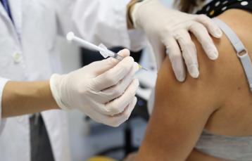
Neonatal Surgery is the sub-discipline within paediatric surgery that deals with surgically treating congenital and acquired illnesses in newborns and infants up to one month old.
This is a highly complex sub-speciality that only exists at tertiary paediatric centres. The past few decades have brought about important advances, thanks to improvements in diagnostic techniques, neonatal intensive care and anaesthetics, and surgical techniques and materials. These have radically changed the prognosis for both birth defects and acquired surgical pathologies in newborns.
Description
Neonatal surgery requires detailed knowledge of complex pathologies in patients who also have special conditions that are different from other paediatric patients. The Neonatal and Foetal Surgery Unit covers practically every surgical neonatal pathology, and it is a reference centre both nationally and on a European level. It is part of ERNICA, the European Reference Network (ERN) for Rare Inherited and Congenital Digestive Disorders. In recent years, the Unit has been firmly committed to introducing minimally invasive surgical techniques, achieving excellent results.
We work in conjunction with the Neonatology Department. The Neonatal Intensive Care Unit (NICU) at Vall d’Hebron Hospital is one of the largest in the country and has one of the best survival rates. We have the possibility of performing surgical interventions within the NICU if transporting the newborn patient to the surgical wing would be too risky due to the instability of their condition.
Similarly, we work closely with the Foetal Medicine Unit, whose services include all the currently valid prenatal and intrapartum (EXIT technique) foetal surgery techniques.
The Unit participates in the following sessions and committees: Neonatal Medical-Surgical Session (Tuesdays at 1 pm), Paediatric Airway Committee, Birth Defects Committee.
Within our areas of interest, we highlight oesophageal atresia, a pathology for which our overall survival rate exceeds 95% in patients who do not present complex associated malformations. Patients with long-gap oesophageal atresia continue to be a challenge, as the distance between the ends is so great that a primary repair cannot be done. In these complex patients, besides applying the different classic techniques, we use the Foker technique, which consists of sustained traction on the ends to be connected in order to stimulate their growth, so that oesophageal anastomosis can finally be achieved. In those patients for whom the native oesophagus cannot be saved, we carry out oesophageal substitution via gastric pull-up, with excellent short-term and long-term results.
We have established a comprehensive treatment plan for patients affected by a congenital diaphragmatic hernia (CDH), which includes foetal treatment (using the FETO technique: Foetal Endoscopic Tracheal Occlusion) in cases where CDH has been diagnosed in the foetus and the prognosis is not good. In addition, the hospital offers every type of invasive and non-invasive respiratory support and therapy available today: synchronised mechanical ventilation, HFOV, CPAP, treatment with inhaled nitrous oxide, and ECMO (extracorporeal membrane oxygenation), providing cardiovascular and respiratory support when conventional treatment proves ineffective. In patients with large diaphragmatic defects, we use several types of patches, both synthetic and biological ones, and techniques that use autologous muscle flaps, generally from the abdominal wall.
The main abdominal wall defects we find are gastroschisis and giant omphalocele. We are especially proud of how we handle gastroschisis, which can be associated with severe medical-surgical problems due to the inflammation and thickening of the exposed intestinal loops caused by the irritation produced by the amniotic fluid at the end of gestation. Since 2002, we have been carrying out a strategy to avoid this, consisting of performing a scheduled Caesarian section at 34-35 weeks of gestation. This elective preterm C-section technique allows for the abdominal defect to be closed directly, as it reduces exposure to amniotic fluid. With more than 50 patients treated this way, we have not observed complications linked to prematurity. Instead, we have detected a decrease in the associated complications, an earlier introduction to food, and a reduced hospital stay, as well as better aesthetic outcomes, since the scar is hidden by the belly button. We have vast experience in treating giant omphalocele. For this, we use surgical techniques that employ vascularised flaps, biologic or synthetic mesh, and vacuum-assisted closure (V.A.C.®).
Our overall survival rates for acquired surgical pathologies such as necrotising enterocolitis and intestinal perforation (both in premature and LGA premature - with a birth weight of less than 750g - babies) is comparable to other European reference centres. This is thanks to the application of our philosophy of minimally aggressive surgery and conservative treatment; applying these principles is the underlying reason for our high rates of survival and preservation of native intestine.
Portfolio of services
Intrauterine or peripartum surgery
- Abdominal or thoracic malformations or tumours that require prenatal or intrapartum surgical treatment (using the EXIT technique, ex utero intrapartum treatment)
Pathologies of the disgestive tract
- Treatment of necrotising enterocolitis
- Treatment of intestinal perforation
- Surgical treatment of ileus and meconium peritonitis
- Following - Repairing oesophageal atresia, using both thoracoscopic and open approaches We specialise in complex patients (those with associated heart disease) and managing long-gap oesophageal atresia
- Oesophageal lengthening techniques to repair long-gap oesophageal atresia (the Foker technique), using both thoracoscopic and open approaches
- Oesophageal substitution via gastric pull-up, using minimally invasive and open approaches
- Congenital or acquired oesophageal stenosis, endoscopic or surgical treatment
- Gastrostomy feeding tube (open or percutaneous endoscopic techniques)
- Treatment for gastric perforation and volvulus
- Hypertrophic pyloric stenosis, pyloric and prepyloric atresia, using minimally invasive and open approaches
- Repairing congenital or acquired intestinal atresia and stenosis, using minimally invasive and open approaches
- Repairing duodenal atresia and annular pancreas, using minimally invasive and open approaches
- Treatment for intestinal duplication, using minimally invasive and open approaches
- Treatment for Meckel's diverticulum and persistent omphalomesenteric duct, using minimally invasive and open approaches
- Repairing defects involving intestinal rotation and volvulus, using minimally invasive and open approaches
Pathologies of the abdominal wall
- Repairing inguinal hernias in newborns
- Repairing gastroschisis (primary closure, Schuster technique)
- Repairing omphalocele and umbilical cord hernias
- Surgical separation of conjoined (“Siamese”) twins: joined at the thorax, abdomen, or pelvis
Pathologies of the lungs and respiratory tract
- Repairing congenital and acquired tracheoesophageal fistulas (through surgery, endoscopy, and lasers)
- Repairing congenital diaphragmatic hernia in newborns, using minimally invasive and open approaches
- Repairing diaphragmatic paralysis and/or eventration, using minimally invasive and open approaches
- Tracheostomy
- Rigid and flexible bronchoscopy
- Surgical treatment of complicated congenital pulmonary pathologies in newborns (cystic adenomatoid malformation, bronchopulmonary sequestration, CPAM, congenital lobar emphysema)
- Treatment of congenital chylothorax
Others
- Treatment for vascular malformations (including complex lymphatic malformations of the head and neck), in coordination with the Paediatric Vascular Anomalies Committee and the Foetal Surgery Unit
- Cervical teratoma and epignathus
- Treatment for intrauterine and post-natal testicular torsion
- Excision of sacrococcygeal teratoma, in coordination with the Oncology Surgery Unit and the Foetal Surgery Unit
- Temporary or permanent vascular access points


")


")

Guillén Burrieza


Castillo Salinas
Suy Franch
Carreras Moratonas
Vazquez Mendez
Herrero García
Arévalo Martínez
Bueno Recio
Ruiz Campillo
Gutiérrez Barceló
Catalán Martínez
García Manau
Goya Canino
Brik


")


