Foetal Medicine and Surgery Unit
The Foetal Medicine and Surgery Unit monitors pregnancies in which the future baby will require special care, such as in cases of multiple births or twins, or when the future baby has a health problem. We are specialists in intrauterine surgery, and our experience makes us a reference point in Catalonia, Spain and around the world.
Description
In the Foetal Medicine and Surgery Unit, we use cutting edge technology to treat the future baby and the mother.
We provide personalised care for the mother, monitor her pregnancy and offer her the most appropriate treatment. We also work alongside professionals from other specialist areas, assessing the mother together and ensuring the health of the future baby before it is born.
We have an Intensive Medicine and Intermediate Obstetric Care Department that works to address any possible health complications in the women. With regards to the future baby, we perform diagnostics with cutting edge technology and offer intrauterine treatments, with the help of some of the leading neonatology units in Spain.
Types of pregnancies and complications treated by this Unit:
- Pregnancy with malformations of the future baby: cardiac, neurological, pulmonary, abdominal, etc.
- Multiple birth: twins, triplets, quadruplets, etc.
- Complications in monochorionic pregnancy (pregnancy with twins with a single placenta): selective intrauterine growth restriction, foetal transfusion, TRAP sequence, etc.
- Maternal infections that could affect the future baby: cytomegalovirus, parvovirus, toxoplasmosis, Zika virus, etc.
We also perform all kinds of foetal surgeries, with the Unit being a figurehead in the fetoscopic treatment of spina bifida, congenital diaphragmatic hernia, monochorionic gestation, foetal valvuloplasty, etc.
Disorders related to this speciality
- Foetal spina bifida
- Pathology associated with the monochorionic gestation: fetofetal transfusion (FFT), selective growth restriction, twin anaemia polycythemia sequence (TAPS).
- Cerebral abnormalities (central nervous system)
- Facial abnormalities: facial clefts (lip and/or palate), micrognathia, anophthalmia/microphthalmia, congenital cataracts.
- Congenital heart disease
- Diaphragmatic hernia
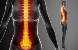
- Spinal abnormalities (hemivertebrae, scoliosis)
- Thoracic masses (pulmonary sequestration, congenital malformation of the airway)
- Abdominal wall abnormalities (omphalocele, gastroschisis)
- Digestive system abnormalities (abdominal cysts, digestive system obstructions, hyperechogenic bowel)
- Urinary tract dilation
- Polycystic kidney disease
- Skeletal abnormalities: clubfoot, polydactyly, skeletal dysplasia
- Genital abnormalities: hypospadias, cryptorchidism
- Genetic syndromes
Treatments related to this speciality
- Prenatal spina bifida surgery
- Clotting of placental vascular anastomoses for fetofetal transfusion
- Cord occlusion in complicated monochorionic pregnancies
- Fetoscopic endoluminal tracheal occlusion (FETO) for congenital diaphragmatic hernia
- Ex-utero Intrapartum Treatment (EXIT)
- Amniotic band resections
- Placement of foetal bypass valve or shunt
- Intrauterine transfusion
- Foetal heart surgery (valvuloplasty)
Diagnostic tests associated with this speciality
- Foetal echocardiogram: this procedure is carried out when it is suspected, through a basic ultrasound scan, that the future baby may suffer from a heart malformation, or if any malformations occurred in a previous pregnancy. It is performed via the abdomen, just like any other ultrasound scan. This procedure generally takes place after 15 weeks of the pregnancy. It is recommended if the future baby is not developing normally in the uterus, or if the mother is taking certain drugs or suffers from any disorders that may increase the risk of congenital heart disease.
- Foetal neurosonography: this procedure is carried out when it is suspected, through a basic ultrasound scan, that there may be an abnormality in the brain of the future baby, or if any cerebral malformations have occurred in previous pregnancies. It is performed via the abdomen, just like any other ultrasound scan. If the future baby is upside down, the procedure can be carried out through the vagina.
- Amniocentesis: this consists of obtaining a sample of amniotic fluid in order to perform different studies (genetic, infection or metabolic). The mother's abdomen is punctured using ultrasound control in order to access the amniotic cavity and obtain a small sample of amniotic fluid.
- 3D/4D ultrasound: a 3D ultrasound is used to create a reconstruction of part of the future baby’s body, allowing for it to be studied in greater depth. A 4D ultrasound adds movement to the 3D images, offering a more complete perspective of the body part.
- Foetal MRI: this test helps us to study certain abnormalities that have been detected in an ultrasound scan.
Departments associated with this speciality
- Obstetrics and Reproductive Medicine
- Clinical and Molecular Genetics
- Neonatology
- Paediatric Radiology
- Paediatric Surgery
- Neonatal Surgery
All paediatric specialities: maxillofacial surgery, urology, child orthopaedics, neurosurgery, ophthalmology, neurology and radiodiagnosis.
